Course Content
Fluid Resuscitation in Sepsis
Course Content (Viewing Only)
The Importance of Fluid Resuscitation in Sepsis
- In patients with septic shock, fluid resuscitation is a critical intervention that restores tissue perfusion. However, there is a great deal of variation in the type of fluid, rate of administration, and the total volume of fluid administered.
- IV fluids should be prescribed like any other drug we give our patients: with a critical look at the indication and contraindication for different types, while balancing the risk of administering too little vs the increasingly apparent sequelae of over administering (1)
- Circulatory insufficiency and shock result from inadequate perfusion relative to the tissue demands (2). Often times, the demand side of perfusion imbalance is an overlooked means of reversing the pathophysiology of shock. Early goal-oriented fluid resuscitation may be crucial in deciding the outcome, but it is important to be aware that the pathogenic inability of the mitochondria to utilize available oxygen may limit the effectiveness of hemodynamic interventions.
- What can cause circulatory insufficiency? The three basic contributors are pump failure, insufficient vascular tone (the vasodilation we see in sepsis), and hypovolemia (2).
- Patients with distributive shock (the primary shock seen in sepsis) may experience circulatory insufficiency due to the profound vasodilatation that is associated with the inflammatory reaction to an infection. (3). As shock progresses and the mean arterial pressure (MAP) trends downward, the cerebrocortical functions are the first to be impaired, which often manifests as a change in the level of consciousness or altered mental status (2).
- As perfusion decreases from vasodilation or from decreased cardiac function, tissue beds become globally hypoxic. The GI tract and the kidneys are known to be intolerant of hypotension and precipitous decrease in MAP, which can lead to impaired mucosal function, bowel integrity, or in extreme cases frank ischemic necrosis (2).
- Secondary to developing tissue hypoxia, and as a result of anaerobic metabolism in the absence of adequate tissue oxygenation, patients may develop a high lactic acid level as a response to the progressive tissue hypoxia. A serum lactate of 4 or greater is associated with increased severity of illness and poorer outcomes even if hypotension is not present (3).
- Knowing this, providers should be prepared to fluid resuscitate patients who are hypotensive or have a lactate >/= 4 mmol/L in order to expand their circulating blood volume and restore tissue perfusion pressure (3).
The Great Debate: Crystalloid vs. Colloid
- Crystalloids are the low-cost salt solutions that are known to be the go-to, easy to grab, and often time’s first choice fluids (4). Isotonic crystalloids are the most commonly administered IV fluid internationally- fun fact: crystalloid solutions were first prepared in response to the cholera pandemic in 1832. So they’ve been around since forever.
- Crystalloids evolved over the next century into two broad groups: sodium chloride (ex. Normal saline) and physiologically-balanced solutions (ex. Lactated Ringers or Plasmalyte). Only about 20-30% of administered crystalloid fluid will stay in the intravascular space. (5).
- Colloids are suspensions of molecules in a carrier fluid with high enough molecular weight to prevent crossing healthy capillary membranes, thus a larger percentage of the administered fluid will remain intravascular. (5)
- Colloids are more expensive fluids and are either man-made (starches, dextrans or gelatins) or naturally occurring (albumin or fresh frozen plasma) (4).
- The physiologic rationale behind favoring colloid over crystalloid is the thought that colloids may expand intravascular volume more effectively by remaining in the intravascular space and maintaining colloid oncotic pressure (5).
- The use of human albumin was first introduced during WWII for victims of traumatic or thermal injuries. Interesting- but a bit cringe-worthy to think of the sterility of this WWII human albumin…
- In the Saline versus Albumin Fluid Evaluation (SAFE) study, a prospective, controlled, randomized, double-blind study comparing 4% human albumin with 0.9% normal saline solution in critically ill patients requiring fluid resuscitation, there was near identical mortality rates in both patient groups. However, there was a small, statistically nonsignificant benefit to using human albumin in the sepsis subgroup. The current SSC guidelines have a weak recommendation (2C) for the administration of albumin in patients with severe sepsis or septic shock who are receiving large amounts of crystalloid fluid. (3).
- In 2013, the Colloids versus Crystalloids for the Resuscitation of the Critically Ill (CRISTAL) study, researchers investigated mortality of ICU patients resuscitated with crystalloids versus colloids (6). After 28 days, there was no statistically significant difference in mortality between the two groups. However, the study continued to track patients through day 90 and found fewer deaths in the colloid group as compared to the crystalloid group (30.7% vs 34.2%, p= 0.03) (6). The colloid group also experienced fewer days of mechanical ventilation and fewer days needing vasopressor therapy (6). While these results seem to be impressive, the study used isotonic crystalloid fluid in both patient groups as maintenance IV fluid, and many patients were treated with different fluids that randomized to prior to study enrollment. Thus, these results should be used with caution given the major study design flaws.
- (The Albumin Italian Outcome Sepsis (ALBIOS) trial in 2014 was a large RCT that compared the administration of crystalloid to the administration of 20% albumin with crystalloid (6). At 28 days and at 90 days there was no significant change in mortality between the two groups (6). At 28 days, 31.8% of patients in the albumin group vs 32% of patients in the crystalloid group had died (p=0.94) (6). Similarly, at 90 days 41.1% of the albumin group and 43.6% of the crystalloid group had died (p=0.29) (6).
- The results of the SAFE, CRISTAL, and ALBIOS trials did not show a clear benefit associated with the use of albumin and combined with the increased cost, crystalloids are the fluid of choice for patients with a diagnosis of sepsis. As research continues, we may begin to see a trend towards better outcomes with albumin administration, but it is currently not recommended as first-line treatment.
Learner exercise
 How will you incorporate the evidence presented in the above information in your practice?
In what types of situations would you use crystalloids VS colloids?
How does the cost of colloid factor into the decision making process, especially when weighed against the negligible potential difference in outcomes?
Choice of Crystalloid Fluid for Resuscitation: Is Normal Saline Really “Normal”?
- The use of saline versus a balanced crystalloid solution has been a topic of ongoing debate.
- Due to the high mortality, the burden to society, and increasing awareness surrounding sepsis, there has been extensive research done to identify the optimal fluid treatment protocols. Ultimately, much of the choice is dictated by provider preference, hospital protocol, and regional availability due to a lack of evidence-based guidelines on specific fluid choice.
- Fluid composition can have unintended physiological effects, such as altering the pH balance through the metabolism of lactate and acetate leading to a decrease in bicarbonate, and eventually metabolic acidosis, and the potential for acute kidney injury (6).
- The Saline Against Lactated Ringers or Plasma-Lyte in the Emergency Department (SALT-ED) study is a pragmatic, cluster, multiple-crossover trial at a single center evaluating the clinical outcomes of patients treated with 0.9% NS versus balanced crystalloids in the setting of resuscitation in the emergency department (7). The trial included 13,347 patients who received a median of 1 liter of fluid (7). Saline increased the risk of death or renal failure when compared to LR/Plasmalyte (5.6% vs 4.7%, p= 0.02). The subgroup of patients with renal injury at the time of admission was more susceptible to adverse kidney events from saline administration (37.6% vs 28%, p= <0.001) (7). This trial confirmed that saline increases the risk of renal failure when compared to balanced solutions.
- These results were then duplicated at Vanderbilt and included critically ill patients. The SMART trial, a pragmatic, cluster-randomized, multiple – crossover trial, was conducted in five intensive care units at an academic center and included 15,802 adult patients (5). Patients were randomized to receive either 0.9% NS or LR/Plasmalyte. Among the 7,942 patients in the balance crystalloid group, 1,139 (14.3%) had an adverse kidney event, compared to 1,211 of the 7,860 (15.4%) patients in the NS group (p = 0.04) (5). In-hospital mortality at 30 days was higher in the NS group when compared to the balanced crystalloid group, as well (11.1% vs 10.3%, respectively, p = 0.06) (5). The mortality difference in the two groups suggests that NS may not only be causing renal failure but may also be causing harm to patients via additional mechanisms, including increased inflammation. (8).
- Normal saline as a resuscitation fluid should not be administered in high amounts as it carries the risk of inducing a hypernatremic hyperchloremic metabolic acidosis (8). Some patients are already extremely acidotic, and by giving them fluid that will exacerbate their academia is poor practice. The truth is that “normal” saline is not physiologically normal. It is a hypertonic, acidotic fluid that can cause more harm than good, especially in patients who need large volume resuscitation. That being said, the development of electrolyte disturbances secondary to fluid administration also depends on the electrolyte status of the patient before resuscitation is initiated.
- In an RCT of patients with pancreatitis that included a comparison between LR and NSS, patients who were treated with NS had higher levels of C-reactive protein one day after the initiation of resuscitation (p = <0.05), meaning that increased saline administration may have a direct correlation to the degree of inflammation (8).
- All of this is not to say that saline is all bad and should never be used, but to point out that just because “it’s what we’ve always used”, “it’s easy to grab”, or “the patient is hyperkalemic!” are not justifiable reasons to use high volumes of NS in the resuscitation of your patients. There are better options, and believe it or not, hyperkalemia is not a contraindication to LR! One liter of LR only contains 4 mEq of potassium!
- While not all balanced crystalloids are created equal, the SALT-ED trial provides good evidence that LR is superior over NS with a number needed to treat (NNT) of 111, which is currently the highest evidentiary support for any balance crystalloid (7).
Learner exercise
Balanced crystalloids may have an advantage over saline-based solution for IV fluid resuscitation. How will you incorporate this into your practice?
What types of patients are likely to benefit from a saline-based resuscitation VS balanced crystalloids?
Some of the unintended effects of saline administration can be high-priority, such as renal failure and a higher risk of death according to some studies. Should we always use balanced solutions when electrolytes permit?
How much fluid do septic patients need?
- The concept of prompt IV fluid administration was first accepted after the 2001 study of early goal-directed therapy (EGDT). In the study, patients either received standard therapy, which involved arterial and central venous catheterization and a protocol targeting a CVP 8-12 mmHg, mean arterial pressure (MAP) at least 65 mmHg, and urine output of at least 0.5 ml/kg/hr, or the EDGT group (5). The EDGT group included the aforementioned components but also included a catheter to measure central venous oxygen saturation (SvO2), six hours of treatment in the emergency department before admission, and administration of 500 mLs of crystalloid fluid every 30 minutes to achieve CVP goals, vasopressors, and vasodilators to maintain MAP goals, and blood transfusions or dobutamine to achieve ScvO2 >/= 70% (5). Overall, in-hospital mortality was found to be 16% less with EDGT when compared to standard therapy (46.5% vs 30.5%; p= 0.009) (5).
- The results of this landmark study propelled early and protocolized fluid management to the forefront of sepsis management. Because of this study and future studies that replicated the above results, the Surviving Sepsis Campaign (SSC) began promoting EGDT fluid resuscitation as a cornerstone of sepsis and septic shock management (5).
- The SSC 3-hour and 1- hour bundle both recommend the initial administration of 30 mL/kg of crystalloid fluid for hypotension or lactate >/= 4 mmol/L as a fluid challenge with a target CVP goal >/= 8 mmHg, ScvO2 of >/= 70%, and normalization of lactate (9). A patient may need repeat fluid challenges in the initial phases of sepsis/septic shock, and this bolus dose is meant to rapidly expand the patient’s blood volume to allow for providers to assess the patient’s response to fluid resuscitation.
- A key concept for dosing fluid therapy in the critically ill population is to actively address ongoing losses (drains, stomas, fistulas, or hyperthermia, open wounds, or various causes of polyuria) paired with the frequent reassessment of the need for further hemodynamic support (10). While fluid administration is a critical aspect of resuscitation, excessive fluid accumulation has been associated with worse clinical outcomes- particularly the development of acute kidney injury (AKI), pulmonary edema, pleural effusions, and in some cases an increase in ventilator days (10).
- The idea of interrelated phases of fluid management, coined “ebb and flow”, differentiated according to the patient’s clinical status with evolving goals for fluid need is highly individualized, but an important concept in the management of the septic patient in order to avoid adverse events related to poor fluid management (10).
- In the initial phase of fluid resuscitation, the objective is the restoration of effective circulating blood volume, organ perfusion, and tissue oxygenation. Fluid accumulation and a positive fluid balance are to be expected here (10). This is basically the only phase we’ve discussed thus far.
- In the second phase, the goal is a maintenance of intravascular volume homeostasis (10). The goal is to prevent excessive fluid accumulation and to avoid unnecessary fluid loading. By the second phase, the patient should show evidence of adequate tissue perfusion.
- In the third and final stage, the objective centers around fluid removal and the concept of “de-resuscitation” as dictated by the state of physiologic stabilization, organ injury recovery, and convalescence (10). During this phase, unnecessary fluid accumulation may add to secondary organ injury and adverse events.
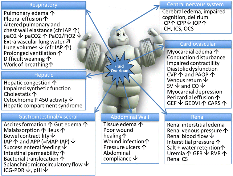
- Below is a photo depicting the potential consequences of fluid overload on end-organ function as adapted by Malbrain et al (1).
Macrocirculation End Points of Sepsis Resuscitation
- As mentioned previously, resuscitation goals for the septic patient are to return the patient to a physiologic state that promotes adequate organ perfusion along with matching metabolic supply and demand.
- Ideally, resuscitation end points should assess the adequacy of tissue oxygen delivery (DO2), oxygen consumption (VO2), and should be quantifiable and reproducible. Since there fails to be a single resuscitation endpoint despite years of research, providers must be able to rely on multiple endpoints in order to determine the patient’s overall response to therapy (11). The SSC focuses their resuscitation guidelines on the original EGDT protocol with an emphasis on macro and microcirculatory endpoints (CVP 8-12mmHg in spontaneously breathing patients/12-15mmHg in ventilated patients, MAP >/= 65mmHg, urine output >/= 0.5mL/kg/hr, central venous oxygen saturation >/= 70% or mixed venous oxygen saturation >/= 65%) (11).
- The ProCESS, ARISE, and ProMISe trials have all compared the original EGDT protocol with contemporary care and have found no difference in clinical outcomes, thus prompting the thought that the one size fits all approach to sepsis may be an outdated approach to treatment.
Learner Exercise
30mL/kg can be a very large amount of fluid in patients with high body weights. Would you still follow the recommendation of 30mL/kg in these cases?
There is much debate about the optimal amount of fluid resuscitation. What are some of the concerns with over and under resuscitation. Which is likely more detrimental in terms of mortality?
Macrocirculation End Points of Sepsis Resuscitation: Central Venous Pressure
- A previously well-established starting point in determining a patient’s need and subsequent responsiveness to fluids is to utilize a static measurement, such as the central venous pressure (CVP) or pulmonary artery occlusion pressure (PAOP) (11). As most providers know- using the CVP as an initial resuscitation target and estimate of preload adequacy is fundamentally flawed. Factors such as total blood volume, cardiac output/venous return, pulmonary hypertension, cardiac tamponade, arrhythmias, and human error involving leveling of the transducer are all factors that have the potential to impact the central venous pressure (11). The correct interpretation of a CVP value is as follows: a low CVP value of </= 6 almost always indicates hypovolemia. However, a high value does not exclude hypovolemia nor does it guarantee hypervolemia.
Microcirculation End Points of Sepsis Resuscitation: Lactate
- Moving from a “macro” point of view to a “micro” point of view, there are several clinical and laboratory values that providers use to assess the microcirculation. Most commonly, lactate, central venous oxygenation, and capillary refill time.
- Lactic acid is one of the most widely accepted biomarkers used to diagnose sepsis-related organ dysfunction. Typically, a lactate >/= 4mmol/L was used as the threshold for organ dysfunction, but recently a threshold of >/= 2mmol/L has been used (11). The working theory behind increased lactate in septic shock is that as global tissue hypoxia occurs, oxygenation fails to meet tissue oxygen demand, therefore increasing anaerobic metabolism…and lactic acid level (11). Just like when you show up for that 1st day of spring 5K after spending the last 4 months on your couch watching Netflix documentaries….need.more.oxygen!!!
- Unfortunately, this very basic explanation fails to consider other contributions to elevated lactate. It continues to be widely accepted and used as a marker of microperfusion, but providers should be aware that there are still limitations.
- Elevated lactate can be attributed to 4 broad categories: decreased tissue oxygen delivery, underlying disease, drugs/toxins, and inborn errors of metabolism (11).
- In decreased oxygen delivery, you could see elevated lactate in individuals who have had a tonic-clonic seizure, severe asthma attack, severe anemia, carbon monoxide poisoning, or chose to do one of those Spartan Races in July. In the underlying disease subset, you could have increased lactate in patients who have fulminant liver failure, lymphoma/leukemia, small cell lung cancer, pheochromocytoma, or thiamine deficiency (sepsis would also fall under this category) (11).
- Drugs and toxins that can often be responsible for an increased lactate include Biguanides, Linezolid, Cyanide poisoning, NRTIs, and beta 2 agonists (11). The rarer inborn metabolism errors are the patients who have enzyme deficiencies such as pyruvate dehydrogenase, pyruvate carboxylase, Fructose-1-6,-diphosphatase, and Phosphoenolpyruvtate carboxykinase (11).
Microcirculation End Points of Sepsis Resuscitation: SvO2/ScvO2
- Moving past lactate measurement to more technical measurements tissue oxygenation, both mixed venous oxygen saturation (SvO2) and ScvO2 have been considered as important targets because they can be used to estimate a global balance of cellular oxygen demand vs delivery (11). A ScvO2 <70% is indicative of inadequate oxygen delivery to tissues, increased oxygen extraction, or a combination of the two. It’s important to note that a true ScvO2 has to be obtained via a central venous catheter with the tip appropriately placed at the junction of the superior vena cava and the right atrium (11).
- Assuming it is measured at the correct location, a ScvO2 of 70%-89% is suggestive of a well-balanced VO2/DO2. A ScvO2 >/= 90% suggests poor oxygenation utilization at the cellular level, tissue dysoxia, or microcellular shunting (11). Currently, the routine use of SvO2 and ScvO2 is not supported in the literature, but the role may become more apparent as sepsis end-goal resuscitation research continues to increase in prevalence.
Microcirculation End Points of Sepsis Resuscitation: Capillary Refill Time
- While technology and invasive tests offer pertinent information, these interventions should be performed in conjunction with frequent clinical examinations to assess the response.
- Capillary refill time is a basic examination skill that new literature is examining as a valuable tool to assess regional and global tissue perfusion during septic shock resuscitation.
- Capillary refill time is defined as the duration of the time needed for the patient’s fingertip to regain color after direct pressure is applied to cause blanching. In a healthy patient, the refill time should be <3.5 seconds (11). It’s important to note that skin temperature, room temperature, age, and use of vasoactive medications can impact capillary refill time and should be taken into consideration. Assuming the patient’s extremities are normothermic, a refill time of >5 seconds suggests the presence of abnormal microcirculatory flow (11).
- Serial assessment with normalization within 6 hours is associated with successful resuscitation when compared against traditional resuscitation targets.
Estimating Fluid Responsiveness with SVV and bedside echocardiography
- Dynamic indices such as stroke volume variation (SVV), pulse pressure variation, and inferior vena cava variability all have been found to have a better predictive value, sensitivity, and specificity than the static indices (11). In patients who are spontaneously breathing or have arrhythmias, direct measurement tests such as the expiratory occlusion test and passive leg raise may be preferred (11).
- As SVV of >12 has an 88% sensitivity and 89% specificity for predicting fluid responsiveness in patient without cardiac arrhythmias and requiring mechanical ventilation. Some monitoring equipment may calculate SVV with a standard arterial line only, other times a special arterial line may need to be inserted in order to measure SVV (15).
- Sepsis-induced cardiac dysfunction is well described and often presents as a reduction in left ventricular stroke volume and impaired myocardial performance. Non-invasive ways to measure the cardiac output and cardiac indexes, such as the FloTrac or Vigileo systems or basic bedside echocardiography, have become more common. The use of invasive pulmonary artery catheters are associated with more risk than patient benefit and their use has significantly decreased. Information gained from bedside echocardiography includes: rough estimate of cardiac output, LV and RV function, chamber fluid status, IVC size and variability, and global cardiac function. This information can be invaluable when utilized in real-time, especially to measure the responsiveness of treatments.
Learner exercise
There are multiple types endpoints we can use to measure fluid resuscitation and volume status. Which end points are favored in your clinical practice?
As an extension to the previous question- will you incorporate any of the information gained in this module into your practice?
Fluid challenge without the fluid: Passive leg raise and end-expiration occlusion test
- What could be better than determining the effect of a fluid bolus without actually infusing any fluid? Though these techniques are imperfect, they can provide insight into the “fluid responsiveness” of a patient.
- The passive leg raise test is another noninvasive means to assess fluid need by mimicking a fluid bolus. It involves moving a patient from the semi-recumbent position to a position where the legs are lifted at 45 degrees and the trunk remains horizontal (2). This induces a transfer of 250-350 mL of venous blood from the inferior limbs and the splanchnic compartment towards the thoracic and cardiac cavities mimics the increase in cardiac preload induced by fluid infusion. The threshold to define fluid responsiveness with a passive leg raise test is a 10% increase in stroke volume or cardiac output (1). Cardiac output changes can be detected 1-2 minutes after the maneuver is performed using either SVV via a non-invasive technology (Flo-Trac) or by utilizing bedside echocardiography to visualize the changes in cardiac function (12). A positive response may also be noted if blood pressure increases with a decrease in heart rate, though this is less sensitive and specific. Similar to capillary refill, the passive leg raise can be done regardless of arrhythmia or mode of mechanical ventilation (12).
- The end-expiration occlusion test is another fluid responsive test, but specifically for the subset of patients who require mechanical ventilation. The test consists of stopping mechanical ventilation at end expiration for 15 seconds and measuring the changes in cardiac output. By pausing mechanical ventilation there is an increase in cardiac output by stopping the cyclic impediment of venous return that occurs at each ventilator triggered breath. An increase in cardiac output above the threshold of 5% indicates fluid responsiveness.
Putting it together
- The best approach is to use multiple techniques to measure the efficacy of fluid resuscitation. Relying on any single parameters is not ideal practice and may lead to under or over-resuscitation. The best way to use this data is to perform interventions which increase perfusion (usually a fluid bolus in sepsis) and re-measure the end-point. A trend toward better perfusion (lower lactic acid level, faster capillary refill, etc.) indicates a positive response. A negative response can be due to either: 1.) inadequate volume of fluid resuscitation OR 2.) a patient that is no longer fluid responsive. It can be difficult to discern the difference and the passive leg raise or occlusion test may be helpful here. There is no hard and fast rule, but generally it is thought to be better to over-resuscitate than under resuscitate.
- By systematically using this approach, there aim is to properly resuscitate the patient while avoiding the pitfalls of both over and under-resuscitation. End-points should be measured after each intervention.
- For example if you measure a lactic acid level of 8 and note delayed capillary refill on exam, you may determine that fluids will augment cardiac output and increase tissue perfusion. Thus you choose to administer 1L bolus. Once the bolus is complete, you should re-check the lactic acid level and capillary refill. It may not normalize, but there should be improvement.
Summary
In summary, fluid resuscitation in sepsis is a controversial topic. Nurses should utilize a variety of end-points to measure fluid status and perfusion status. Newest evidence is suggesting that LR may have a physiologic benefit over NS and albumin may have role in the resuscitation of septic patients.
Not a member? Sign up here to complete the course and receive your certificate.
References (Bibliography)
1) Malbrain, M. L., Regenmortel, N. V., Saugel, B., Tavernier, B. D., Gaal, P. V., Joannes-Boyau, O., . . . Monnet, X. (2018). Principles of fluid management and stewardship in septic shock: It is time to consider the four D’s and the four phases of fluid therapy. Annals of Intensive Care,8(1), 1-16. doi:10.1186/s13613-018-0402-x
2) Marini, J. J., & Dries, D. J. (2019). Shock and support of the failing circulation. In Critical Care Medicine: The essentials and more(5th ed., pp. 47-67). Philadelphia, PA: Lippincott Williams & Wilkins.
3) 3- Hour Bundle. (n.d.). Retrieved March 21, 2019, from http://www.survivingsepsis.org/SiteCollectionDocuments/Bundle-3-Hour-Step4-Fluids.pdf
4) Lewis, S. R., Pritchard, M. W., Evans, D. W., Butler, A. R., Alderson, P., Smith, A. F., & Roberts, I. (2018, August 3). Colloids or crystalloids for fluid replacement in critically people. Retrieved March 21, 2019, from https://www.cochrane.org/CD000567/INJ_colloids-or-crystalloids-fluid-replacement-critically-people
5) Semler, M. W., & Rice, T. W. (2016). Sepsis Resuscitation: Fluid Choice and Dose. Clinics in chest medicine, 37(2), 241-50.
6) Avila, A. A., Kinberg, E. C., Sherwin, N. K., & Taylor, R. D. (2016). The Use of Fluids in Sepsis. Cureus. doi:10.7759/cureus.528
7) Self, W. H., Semler, M. W., Wanderer, J. P., Ehrenfeld, J. M., Byrne, D. W., Wang, L., Atchison, L., Felbinger, M., Jones, I. D., Russ, S., Shaw, A. D., Bernard, G. R., … Rice, T. W. (2017). Saline versus balanced crystalloids for intravenous fluid therapy in the emergency department: study protocol for a cluster-randomized, multiple-crossover trial. Trials, 18(1), 178. doi:10.1186/s13063-017-1923-6
8) Farkas, J. (2018, February 27). PulmCrit- Get SMART: Nine reasons to quit using normal saline for resuscitation. Retrieved March 21, 2019, from https://emcrit.org/pulmcrit/smart/
9) Levy, M. M., Evans, L. E., & Rhodes, A. (2018). The Surviving Sepsis Campaign Bundle. Critical Care Medicine,46(6), 997-1000. doi:10.1097/ccm.0000000000003119
10) Mcdermid, R. C. (2014). Controversies in fluid therapy: Type, dose and toxicity. World Journal of Critical Care Medicine,3(1), 24-33. doi:10.5492/wjccm.v3.i1.24
11) Greenwood, J. C., & Orloski, C. J. (2017). End Points of Sepsis Resuscitation. Emergency Medicine Clinics of North America,35(1), 93-107. doi:10.1016/j.emc.2016.09.001
12) Boyd, J. H., Sirounis, D., Maizel, J., & Slama, M. (2016). Echocardiography as a guide for fluid management. Critical Care,20(1), 1-7. doi:10.1186/s13054-016-1407-1
13) Byrne, L., & Van Haren, F. (2017). Fluid resuscitation in human sepsis: Time to rewrite history?. Annals of intensive care, 7(1), 4.
14) De Backer, D., & Vincent, J. L. (2018). Should we measure the central venous pressure to guide fluid management? Ten answers to 10 questions. Critical care (London, England), 22(1), 43. doi:10.1186/s13054-018-1959-3
15.) Xavier M., Marik, P., Jean-Louis T. (2016) Prediction of fluid responsiveness: an update. Annals of intensive care. doi: 10.1186/s13613-016-0216-7